The medial collateral ligament (MCL) goes from the femur to the tibia on the medial side of the knee. Injuries to this ligament are common, as they can happen from being hit on the lateral side of the knee. Other causes of an MCL injury include quick stops and turns, jumping and landing in an awkward position, and hyperextending the knee.

Assessment of an MCL sprain is done with a Valgus Stress Test. The video on Assessment and Treatment of Knee Pain shows this and other tests.
Treatment involves applying multidirectional friction to the affected areas for 20 – 30 seconds, followed by eccentric loading of the three muscles that cross the medial side of the knee. The friction helps draw fibroblasts to the area, and the eccentric loading causes the fibroblasts to lay down collagen in the direction of the muscle fibers and the fibers of the ligament. I also would have my client do the eccentric loading with the other leg so that they feel balanced.
The three muscles that cross the medial side of the knee are the Sartorius, Semitendonosis, and Gracilis.
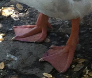
These three muscles cross at the medial side of the knee and attach at the pes anserinus (goose foot) area of the tibia. They help to give stability to the medial side of the knee. These three muscles come from three widely separated origins on the three bones of the innominate (ilium, ischium, and pubis), so they can act as inverted guy wires to stabilize the medial knee. Their common attachment on the medial tibia reminded early anatomists of a goose foot, so they gave it the name pes anserinus, which is Latin for goose foot.


I had to chase a goose around a park to get this picture. 🙂
